NOW ENROLLING
SUMAS Admission
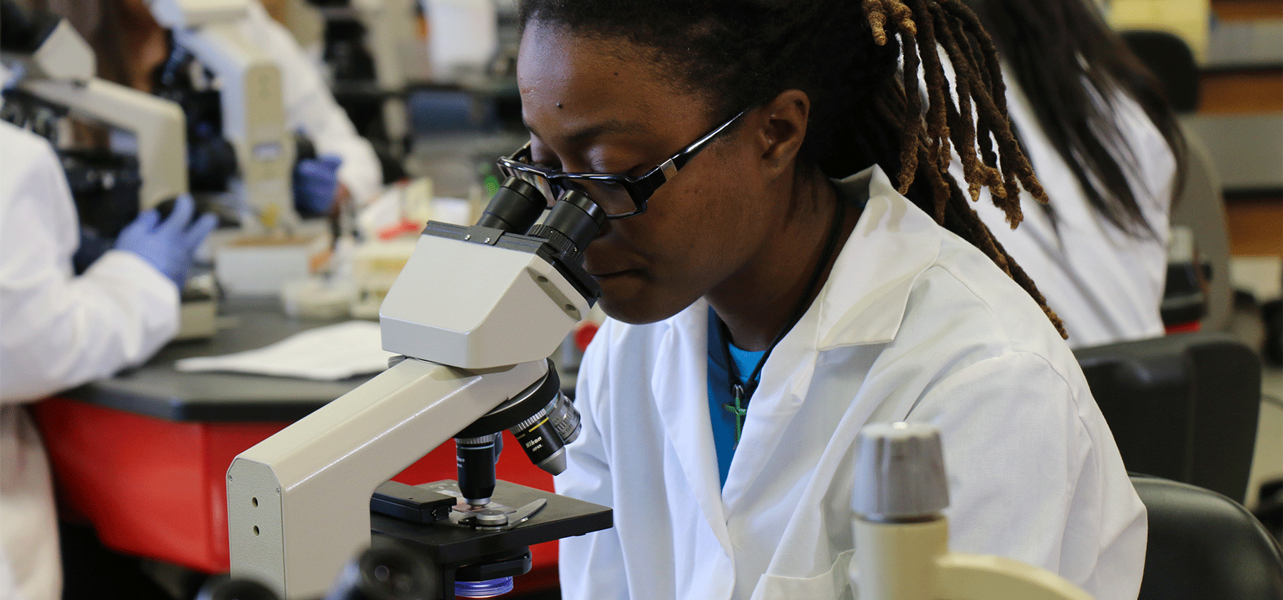
The student is expected to carry out practical exercises in all the disciplines:
Clinical Chemistry: titration, presentation of volumetric analysis. Methods for chloride determination. Determination of bicarbonate in plasma, percentage purity of carbonate. Determination of the composition of the mixture Na0H/Na2C03, NACI/HCI, specific gravity, reactions with ferric chloride, urobilinogen, bilirubin, indicant, myoglobin, cysteine, protein, Bence-Jones protein, blood, reducing substances, ketone bodies, phenyl pyruvic acid. Spectroscopy of plasma and urine CSF analysis – sugar, protein.
Haematology and BGS: Blood film, WBC count, Hb estimation, Absolute values, Eosinophil count. Rectics count. Osmotic Fragility. Blood grouping techniques. Antiserum titration, Anti-human globulin (AHG) direct and indirect, Antibody screening. Donor screening, secretor status.
Histopathology: Preparation of fixatives, removal of formalin pigments, testing of end point of decalcification using chemical methods. General tissue staining by Hematoxylin and counter-staining with eosin. Demonstration of elastic and collagen fibres. Prussian blue (Perl’s) reaction for iron in tissues. Gram and Ziehl Neelsen (Zn) staining methods. Use of automatic tissue processors Microtome.
Medical Microbiology and Parasitology: Safety precautions in the Microbiology laboratory. Getting acquainted with basic tools of microbiologist. Preparation of films and basic staining techniques, the Gram stain, Ziehl Neelsen stain, spores, capsule and negative staining procedures. Wet preparation and microscopy, Motility tests, Media preparation and culturing. Plate reading; demonstration of the ubiquity of micro-organisms especially bacteria from different environment. Recognition of different types of haemolysis. Sensitivity testing. Use of autoclave. Wet mount for parasites. Identification of trophozoites cysts and ova of different protozoa and helminths in stool. Thin and thick films preparation for malaria microfilaria and Trypanosome parasites. Staining techniques: Giemsa, Wrights, Fields and Leishman stains. Identification of Trichomomonas Spp, paragonimus. Trichuris and other helminths and protozoa of medical importance including Schistosomes. Skin snips. Urine microscopy. Concentration techniques for stool and sputum for ova and cysts. Examination and recognition of helminths from tissue Biopsy.